
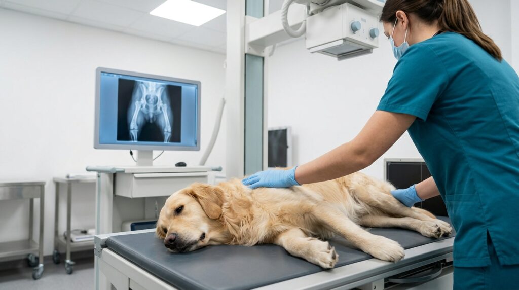

When your veterinarian recommends an X-ray, it can be hard to know what to expect or why it is needed. As a trusted vet near you at South Etobicoke Animal Hospital, we use digital X-rays every day to look past the surface and understand what is really going on inside your pet’s body. This guide breaks down what pet X-rays actually show, when your vet is likely to recommend one, how the appointment works, and how X-rays compare to other diagnostic tools.
What Is a Pet X-Ray and How Does It Work
A pet X-ray, also called a radiograph, uses a small, controlled burst of electromagnetic radiation to create a two-dimensional image of the inside of your pet’s body. Dense structures like bone absorb more radiation and appear white on the image, while softer tissues such as muscle, fat, and gas-filled organs appear in varying shades of gray, allowing your veterinarian to distinguish between different structures at a glance.
Why Digital X-Rays Are the Modern Standard
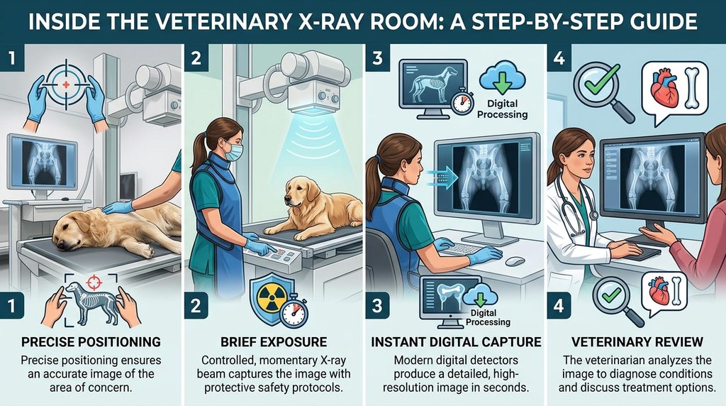
Most veterinary clinics, including ours, now use digital radiography rather than traditional film. Digital systems capture the image electronically, so it appears on screen within seconds rather than requiring darkroom processing. This also means the image can be zoomed, brightened, or contrast-adjusted after the fact, often revealing details that would be missed on a standard film X-ray, and it typically requires a lower radiation dose to achieve a clear picture.
What Conditions Can a Pet X-Ray Detect
X-rays are especially good at showing anything with a different density than the surrounding tissue. Common findings include:
- Bone fractures, dislocations, or signs of arthritis
- Swallowed foreign objects such as toys, bones, or fabric
- Bladder or kidney stones
- Changes in heart size or shape that suggest cardiac disease
- Lung patterns consistent with pneumonia, fluid, or masses
- Spinal abnormalities and disc disease
- Certain tumors, particularly in bone or the chest cavity
Not every condition shows up clearly on an X-ray. Soft tissue detail, such as the internal structure of an organ, is often better evaluated with ultrasound, which is why your veterinarian may recommend both tools depending on the symptoms.
When Your Veterinarian May Recommend an X-Ray
Your veterinary team typically recommends imaging when a physical exam and history raise questions that can only be answered by looking inside the body. Common scenarios include:
- Sudden lameness, limping, or reluctance to bear weight
- A witnessed or suspected foreign object ingestion
- Persistent coughing, labored breathing, or exercise intolerance
- Trauma from a fall, car accident, or fight with another animal
- Straining to urinate or visible blood in the urine
- Pre-anesthetic screening before surgery, to check heart and lung status beforehand
If your pet has an upcoming procedure, imaging is often part of the standard workup described in our guide on how to prepare your pet for surgery, since it helps the surgical team confirm your pet is a safe candidate for anesthesia. For families searching for medical imaging near me, our in-house digital radiography means these scans can usually be completed the same day, without a referral to an outside facility.
X-Ray vs Other Imaging Tools: Which One Fits the Job
Veterinarians often choose between X-ray, ultrasound, and occasionally a referral for CT or MRI, depending on what needs to be seen.
| Imaging Type | Best For | Sedation Typically Needed |
|---|---|---|
| Digital X-ray | Bones, foreign objects, lungs, general body cavity screening | Rarely, unless the pet is in pain or anxious |
| Ultrasound | Soft tissue detail, organ structure, pregnancy confirmation | No, most pets tolerate it awake |
| CT or MRI (referral) | Complex neurological or detailed soft tissue cases | Usually yes, general anesthesia required |
Because X-ray and ultrasound each reveal different information, our team frequently uses them together to build a complete picture. If you want a deeper look at how these two tools compare and when each is used, our full guide to veterinary ultrasound and radiology covers that comparison in detail.
What to Expect During Your Pet’s X-Ray Appointment
Before the Appointment
In most cases, no special preparation is required for a routine X-ray. If your pet needs mild sedation because they are anxious, in pain, or need to hold a difficult position, your veterinarian will discuss this with you in advance and explain what to expect.
During the X-Ray
Your pet will be gently positioned on a padded table, and a technician may use foam wedges or sandbags to help hold the correct position. The actual exposure takes only a fraction of a second, and most studies involve two or three views from different angles to give the veterinarian a complete look at the area of concern.
After the X-Ray: Reviewing Results With Your Vet
Because the images are digital, your veterinarian can usually review them with you within minutes of the scan. They will point out anything abnormal, explain what it means for your pet’s treatment plan, and answer any questions you have before you leave.
Is X-Ray Radiation Safe for My Pet
Modern digital radiography uses a very low, carefully controlled radiation dose, and a single diagnostic X-ray poses minimal risk to your pet. Veterinary teams also follow strict safety protocols, including shielding and proper technique, to keep exposure as low as reasonably possible while still capturing a clear, diagnostic-quality image. According to the Merck Veterinary Manual, obtaining a quality radiograph depends on correct positioning and technique, which is why experienced staff and modern equipment both matter for getting an accurate result on the first attempt.
A Case Example: Catching a Hidden Problem Early
Many pet owners come to us after noticing something subtle, like a dog favoring one leg after a walk or a cat that seems slightly less active than usual. In a typical scenario, an X-ray reveals an early, hairline fracture or a mild change in joint spacing that would not have been obvious on physical exam alone. Because the issue is caught early, treatment can begin immediately, often preventing a minor problem from becoming a more serious, harder-to-treat condition. This is the core value of diagnostic imaging: it turns guesswork into a clear, evidence-based plan.
For broader context on how imaging fits into overall veterinary practice, the American Veterinary Medical Association notes that regular diagnostic screening is a key part of preventive pet healthcare, while institutions such as the Cornell University Hospital for Animals Imaging Service highlight how radiography remains one of the most widely used tools for evaluating a wide range of species and conditions.
Common Myths About Pet X-Rays, Debunked
Pet owners often hesitate before agreeing to imaging, usually because of a few persistent misconceptions. Clearing these up can make the decision easier.
Myth: X-rays are always painful for pets. The exposure itself involves no sensation at all, since your pet simply needs to hold still for a moment. Any discomfort comes from an existing injury, not from the imaging process, and your veterinary team will always handle a painful pet gently, positioning them in the way that causes the least distress.
Myth: One X-ray means significant radiation exposure. A single diagnostic X-ray uses a very small, tightly controlled dose, comparable to routine imaging used in human medicine. Veterinary equipment is calibrated to capture a clear image using the lowest dose necessary, and the exposure itself lasts only a fraction of a second.
Myth: X-rays can diagnose every condition. As covered earlier, X-rays excel at showing bone and density differences but are limited for fine soft tissue detail. A normal X-ray does not always rule out a problem entirely, which is why your veterinarian may recommend additional testing, such as bloodwork or ultrasound, if symptoms persist despite unremarkable imaging.
Myth: Older pets should avoid X-rays because of their age. Age alone is not a reason to avoid imaging. In fact, senior pets often benefit the most from X-rays, since conditions like arthritis, organ enlargement, and certain cancers become more common with age, and imaging is one of the most direct ways to catch these changes early, when treatment options are broadest.
Myth: If my pet seems fine, imaging is not necessary. Pets are skilled at masking discomfort, particularly cats and stoic dog breeds. A pet that seems outwardly normal can still have an underlying issue, such as early arthritis or a slow-growing mass, that has not yet caused obvious symptoms. This is part of why imaging is sometimes included in routine senior wellness workups rather than only being used after a problem appears.
Frequently Asked Questions About Pet X-Rays in Etobicoke
-
Do pets need to be sedated for an X-ray?
Most pets do not need sedation for a routine X-ray, especially if they can hold still comfortably for a few seconds. However, if your pet is in significant pain, anxious, or the study requires an especially precise position, your veterinarian may recommend light sedation to keep them calm, comfortable, and to ensure the images are clear enough for an accurate diagnosis.
-
How long does a pet X-ray take?
The actual X-ray exposure is nearly instant, but a full appointment, including positioning, taking multiple views, and reviewing results with your veterinarian, usually takes between fifteen and thirty minutes. Emergency cases or pets requiring sedation may take a little longer, but digital imaging means there is no waiting for film to develop.
-
Can X-rays show soft tissue problems like tumors in organs?
X-rays are best at showing structures with clear density differences, such as bone, lungs, and some masses, but they are limited when it comes to fine soft tissue detail inside organs. For those cases, your veterinarian will often recommend an ultrasound alongside or instead of an X-ray to get a more detailed, real-time view of the internal organ structure.
-
Is it safe to have my pet X-rayed multiple times?
Occasional diagnostic X-rays, even several over a pet’s lifetime, carry minimal risk because each exposure uses a very small, carefully controlled radiation dose. Veterinary teams only recommend imaging when it will meaningfully guide diagnosis or treatment, and modern digital systems are designed to achieve clear results with the lowest dose necessary.
-
What is the difference between a digital X-ray and a traditional film X-ray?
Digital X-rays capture the image electronically and display it on a screen within seconds, while traditional film X-rays require chemical processing in a darkroom and take longer to produce. Digital imaging also allows the image to be enhanced, zoomed, or adjusted for contrast after the fact, which often helps reveal details that might be missed on a standard film image.
-
Will my pet need to fast before an X-ray?
For most routine X-rays, fasting is not required since your pet stays awake and alert throughout. If sedation is needed, your veterinary team will give you specific fasting instructions ahead of time, since an empty stomach helps reduce the risk of complications during sedation.
Every internal question deserves a clear answer, and a good X-ray is often the fastest way to get one.