When your dog suddenly stops eating or your cat starts hiding more than usual, the challenge is figuring out what is actually wrong. Animals cannot describe their symptoms, and many serious conditions develop internally without visible warning signs. That is exactly where veterinary ultrasound and radiology make a real difference. These imaging tools allow veterinarians to look inside your pet’s body without a single incision, giving them the visual information they need to make accurate diagnoses and start targeted treatment.
At South Etobicoke Animal Hospital, medical imaging is a core part of how our team evaluates and treats patients. Whether it is a routine abdominal scan during a checkup or a rapid X-ray during a late-night emergency, having these capabilities on-site means faster answers and better outcomes for the animals we care for.
What Is Veterinary Medical Imaging and Why Does It Matter?
Medical imaging is a broad term that covers several technologies used to visualize the inside of an animal’s body. In veterinary medicine, the two most commonly used modalities in general practice are ultrasound and digital radiography, which is commonly known as X-ray. Each one serves a different purpose, and veterinarians often use both together to build a complete diagnostic picture.
The real value of imaging lies in its ability to reveal conditions that physical examination alone cannot detect. A veterinarian can palpate the abdomen and feel for abnormalities, but they cannot see the size of a kidney, identify fluid accumulation around the lungs, or spot a small tumour growing on the spleen just by touch. Medical imaging fills that gap. According to the Merck Veterinary Manual, radiography remains the most widely used imaging method in veterinary clinics, while ultrasound excels at providing detailed views of soft tissue structures in real time.
For pet owners searching for an ultrasound clinic or radiology near me in the Etobicoke area, understanding what these tools do and when they are needed can take a lot of the uncertainty out of a diagnostic appointment.
The Role of Diagnostic Imaging in Modern Animal Health Services
Diagnostic imaging is not a standalone test. It works alongside bloodwork, urinalysis, and physical examination to form a comprehensive clinical picture. When your veterinarian recommends imaging, it is usually because the physical exam and initial lab work have raised questions that only a visual look inside the body can answer.
At our clinic, in-house medical imaging is available on-site, which means there is no need to send your pet to a separate specialty facility for routine diagnostic scans. This saves time, reduces stress for your animal, and allows the veterinary team to act on results immediately rather than waiting days for an external report.
Understanding Pet Ultrasound — How It Works and What It Reveals
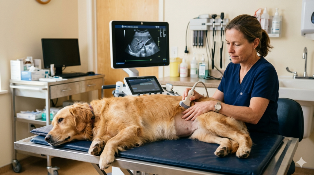
Ultrasound technology uses high-frequency sound waves to create real-time images of internal organs and structures. A small probe, called a transducer, is pressed against the skin and moved across the area being examined. The sound waves bounce off internal tissues and are converted into a visual image on a monitor. There is no radiation involved, no pain for the animal, and in most cases no sedation required.
Pet ultrasound is particularly useful for evaluating soft tissue organs that do not show up well on X-ray. The liver, kidneys, spleen, bladder, adrenal glands, and reproductive organs are all readily visible on ultrasound. It is also commonly used to assess pregnancy, identify masses or cysts, detect free fluid in the abdomen, and evaluate blood flow through major vessels.
Abdominal Ultrasound for Dogs and Cats
Abdominal ultrasound is one of the most frequently requested imaging studies in veterinary practice. It provides a detailed, real-time view of the organs within the abdominal cavity. For dogs, common reasons for abdominal ultrasound include unexplained vomiting, diarrhea, weight loss, changes in urination, abdominal swelling, or abnormal bloodwork that suggests liver or kidney involvement.
For cats, ultrasound is invaluable for identifying conditions like kidney disease, intestinal lymphoma, pancreatitis, and urinary blockages. Because cats are notoriously good at hiding illness, imaging often catches problems that might not be apparent from a physical exam alone.
During the procedure, a small patch of fur may be shaved over the area of interest to improve image quality. The pet lies on a padded table, usually on their back or side, and the veterinarian gently glides the probe across the skin with a water-based gel to eliminate air pockets between the probe and the body. Most dogs and cats tolerate the process well without any sedation.
Soft Tissue Diagnostics With Ultrasound
Beyond the abdomen, ultrasound plays a critical role in evaluating soft tissue injuries and abnormalities. Tendons, ligaments, muscle masses, and superficial lumps can all be assessed with targeted ultrasound scanning. If your dog develops a lump under the skin, ultrasound can help determine whether the mass is solid or fluid-filled, which guides the veterinarian’s recommendation on whether further testing such as a fine-needle aspirate or surgical biopsy is needed.
Echocardiography, which is an ultrasound of the heart, is another important application. It allows veterinarians to assess heart chamber size, wall thickness, valve function, and blood flow patterns. For breeds predisposed to heart disease, echocardiography is an essential screening tool. This kind of advanced veterinary diagnostics helps catch cardiac conditions before they progress to heart failure.
Digital Radiology (X-Ray) — Fast, Accurate, and Non-Invasive
Digital radiography has largely replaced traditional film-based X-ray in modern veterinary practice. The technology works on the same basic principle — a controlled beam of X-rays passes through the body and creates an image based on how different tissues absorb the radiation. Bones appear white because they absorb more X-rays, while air-filled structures like lungs appear dark. Soft tissues fall somewhere in between.
What makes digital radiology superior to older film systems is speed, image quality, and flexibility. Digital images appear on a screen within seconds, can be enhanced or zoomed for closer inspection, and can be easily shared with specialists for second opinions. There is no chemical processing, no waiting, and no repeated exposures due to poor film quality.
Orthopedic Diagnostics With Digital X-Rays
One of the most common uses for X-rays in veterinary medicine is evaluating the musculoskeletal system. Fractures, joint dislocations, bone tumours, arthritis, and hip dysplasia are all conditions that digital radiology identifies clearly. If your dog has been limping after a fall, or your older cat seems reluctant to jump onto furniture, X-rays can quickly reveal whether there is a structural issue that needs treatment.
For pets requiring pet surgery due to a fracture or orthopaedic condition, pre-surgical radiographs are essential for planning the approach. Knowing exactly where and how a bone is broken allows the surgical team to prepare the right equipment and technique before the animal is under anaesthesia. To learn more about surgical preparation and recovery, our guide on common veterinary surgeries for cats and dogs covers the full process in detail.
How X-Rays Detect Internal Abnormalities
Beyond bones, digital radiology is valuable for identifying a wide range of internal conditions. Chest X-rays can reveal heart enlargement, fluid in the lungs, pneumonia, and certain types of cancer that has spread to the thoracic cavity. Abdominal X-rays help detect foreign objects that pets may have swallowed, bladder stones, intestinal obstructions, and abnormal gas patterns that suggest a bowel blockage.
In many cases, X-rays and ultrasound are used together. An X-ray might show that a mass is present in the abdomen, and an ultrasound follow-up provides the detailed soft tissue information needed to characterize it. This combined approach improves diagnostic accuracy and helps the veterinary team move quickly toward the right treatment plan.
Ultrasound vs. X-Ray — Which Imaging Method Does Your Pet Need?
Pet owners often wonder whether their animal needs an ultrasound, an X-ray, or both. The answer depends entirely on what the veterinarian is looking for. Each modality has strengths that the other does not.
X-rays are the preferred choice when the veterinarian suspects bone injury, foreign body ingestion, lung disease, or needs a broad overview of the chest or abdominal cavity. They produce images quickly and provide excellent visualization of dense structures like bone and calcified tissues.
Ultrasound is the better option when the concern involves soft tissue organs, fluid accumulation, cardiac function, or pregnancy evaluation. It provides real-time images that show organ movement and blood flow, which static X-rays cannot capture. Ultrasound is also the preferred tool when guided tissue sampling is needed, such as collecting a biopsy from a liver mass without open surgery.
In practice, most diagnostic workups that involve imaging will include both modalities working together. A dog that presents with sudden abdominal swelling might receive X-rays first to rule out a bowel obstruction, followed by ultrasound to assess the organs and check for internal bleeding. This layered diagnostic approach, supported by in-house diagnostics like bloodwork and urinalysis, ensures that nothing is missed.
Imaging for Emergency Cases — When Every Second Counts
Emergencies are where in-house imaging capabilities make the most dramatic difference. When a pet arrives in critical condition — hit by a vehicle, struggling to breathe, or acutely bloated — waiting hours or days for an external imaging appointment is simply not an option. Rapid access to digital X-rays and ultrasound allows the veterinary team to assess internal injuries, identify bleeding, and determine whether emergency surgery is needed, all within minutes of arrival.
At our emergency vet clinic in Etobicoke, imaging is integrated into the emergency triage process. A dog that has been hit by a car, for example, would receive chest and abdominal radiographs to check for rib fractures, lung contusions, and free fluid. If fluid is detected, a focused ultrasound scan confirms whether there is active internal bleeding. That information determines whether the animal needs immediate surgical intervention or can be stabilized medically.
The speed of digital radiology is a genuine advantage here. Images are available in seconds, not hours. Combined with on-site bloodwork and the clinical judgment of experienced veterinarians, imaging for emergency cases dramatically improves the chances of a positive outcome.
What to Expect During a Pet Imaging Appointment
Knowing what happens during an imaging appointment can help reduce anxiety for both you and your pet. The process is straightforward and, in most cases, completed within a single visit.
Before the Imaging Session
Your veterinarian will discuss the reason for imaging and explain which modality is recommended. If your pet is coming in for a planned ultrasound, you may be asked to withhold food for several hours beforehand. A full stomach can obscure the view of abdominal organs. For X-rays, no specific preparation is usually required unless sedation is anticipated.
As part of a thorough diagnostic workup, your veterinarian may also recommend bloodwork before or alongside imaging. This is where reference laboratory testing comes in. Lab results combined with imaging findings give the most complete picture of your pet’s condition.
During and After the Procedure
X-rays take only a few seconds per image. Your pet is gently positioned on the X-ray table, and the technician steps behind a protective barrier to take the image. Multiple views are usually taken — a side view and a top-down view are standard for most body regions.
Ultrasound takes a bit longer, typically fifteen to thirty minutes depending on the area being scanned. The veterinarian moves the probe slowly across the body, examining each organ systematically. Results are interpreted immediately, and your veterinarian will discuss the findings with you during the same appointment.
Neither X-rays nor ultrasound involve recovery time. Your pet can go home right away, and there are no aftereffects from the imaging itself. As the American College of Veterinary Radiology notes in its consensus guidelines, imaging reports serve as essential documentation that supports clinical decision-making and ongoing patient care.
When Should You Consider Veterinary Imaging for Your Pet?
Imaging is not reserved only for emergencies or seriously ill animals. There are several everyday scenarios where veterinary ultrasound or radiology provides important diagnostic information.
If your pet is due for a wellness and preventive care visit and the veterinarian discovers an abnormal finding during the physical exam — such as a heart murmur, an enlarged organ, or an unusual lump — imaging is often the logical next step. Senior pets, in particular, benefit from periodic imaging as a screening tool. Kidney changes, splenic masses, and early cardiac disease are all detectable through imaging long before outward symptoms appear.
Dental conditions also frequently involve diagnostic imaging. Digital dental X-rays, which fall under a related but distinct category of radiology, are used to evaluate tooth roots and jawbone health below the gumline. If your pet is scheduled for a dental procedure, our guide on cat and dog teeth cleaning explains how dental radiographs fit into that process.
Any unexplained change in your pet’s behaviour, appetite, energy level, or bathroom habits could warrant imaging. Veterinarians rely on these tools to answer the questions that bloodwork and physical exams alone cannot. In the context of comprehensive animal health services, imaging is not a luxury — it is a fundamental part of evidence-based veterinary care.
The Canadian Veterinary Medical Association recognizes diagnostic imaging as a core competency within veterinary practice. Clinics that maintain modern, on-site imaging equipment are better positioned to provide timely, accurate diagnoses and reduce the need for external referrals.
Frequently Asked Questions About Veterinary Ultrasound and Radiology
-
Is veterinary ultrasound safe for dogs and cats?
Yes, pet ultrasound is completely safe. It uses sound waves rather than radiation, making it a non-invasive and painless diagnostic tool. There are no known side effects, and most animals tolerate the procedure comfortably without sedation. Ultrasound is safe for use on pregnant animals as well, which is why it is the standard method for confirming and monitoring pregnancy in both dogs and cats across Etobicoke veterinary practices.
-
What is the difference between ultrasound and digital radiology in veterinary medicine?
Digital radiology uses X-ray beams to create images of dense structures like bones, foreign objects, and the chest cavity. Ultrasound uses sound waves to produce real-time images of soft tissue organs such as the liver, kidneys, and heart. Veterinarians in Etobicoke commonly use both methods together during a single diagnostic workup because each tool reveals different types of information that the other cannot capture on its own.
-
How long does a pet ultrasound or X-ray appointment take?
X-rays are fast — each image takes only seconds, and a complete set of views can be finished in under ten minutes. Ultrasound appointments typically run between fifteen and thirty minutes depending on the complexity of the scan. Results from both modalities are available immediately, allowing your veterinarian to discuss findings and recommended next steps during the same visit without scheduling a separate follow-up appointment.
-
Does my pet need to be sedated for imaging?
Most pets do not require sedation for standard imaging. Ultrasound is gentle and quiet, and the majority of dogs and cats remain calm throughout the scan. X-rays require your pet to hold still briefly, and cooperative animals manage this easily. Sedation may be recommended in specific situations, such as when a pet is extremely anxious, in significant pain, or when precise positioning is necessary for an orthopaedic X-ray series.
-
When should I ask my veterinarian about diagnostic imaging near me?
You should bring up imaging if your pet shows unexplained symptoms like persistent vomiting, sudden weight loss, difficulty breathing, limping, or changes in urination patterns. Your veterinarian may also recommend imaging following abnormal bloodwork results or during senior wellness screening. Early detection through veterinary ultrasound and radiology in Etobicoke improves treatment outcomes and often reduces the overall cost of care by catching conditions before they become advanced.
-
Can imaging detect cancer in pets?
Imaging plays a significant role in cancer detection and staging. X-rays can identify tumours in the chest and visible bone lesions, while ultrasound excels at detecting masses within abdominal organs such as the spleen, liver, and kidneys. Imaging also helps veterinarians determine whether cancer has spread to other areas of the body, which directly influences treatment planning and prognosis for your pet.
-
Does South Etobicoke Animal Hospital offer pet ultrasound near me?
South Etobicoke Animal Hospital provides on-site pet ultrasound and digital radiology as part of our comprehensive diagnostic capabilities. Located at 741 The Queensway in Etobicoke, our clinic serves pet families across Mimico, Long Branch, New Toronto, and the Greater Toronto Area. Our veterinary team performs imaging studies and interprets results the same day, ensuring your pet receives timely and accurate diagnostic care without unnecessary delays or referrals.