

When your veterinarian mentions an ultrasound, it can be hard to know what that actually means for your pet. Is it painful? Does it require anesthesia? What is it actually looking for? At South Etobicoke Animal Hospital, pet ultrasound is one of the most valuable tools available for seeing what is happening beneath the surface without surgery or radiation. This guide breaks down exactly what a pet ultrasound involves, what it can reveal, and the situations where your vet is most likely to recommend one.
What Is a Pet Ultrasound?
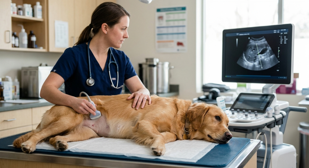
A pet ultrasound is a diagnostic imaging technique that uses high-frequency sound waves to create real-time images of soft tissue and internal organs. A handheld probe is gently pressed against a clipped area of skin, usually the abdomen, and the reflected sound waves are translated into a moving picture on a monitor. Unlike X-rays, which capture a still image, ultrasound shows movement as it happens, which means a veterinarian can watch a heart beating, blood flowing, or a bladder filling in real time.
This makes pet ultrasound especially useful for evaluating soft-tissue structures such as the liver, kidneys, spleen, bladder, intestines, and reproductive organs, areas where a physical exam or bloodwork alone cannot provide a complete picture.
How Pet Ultrasound Differs From X-Rays
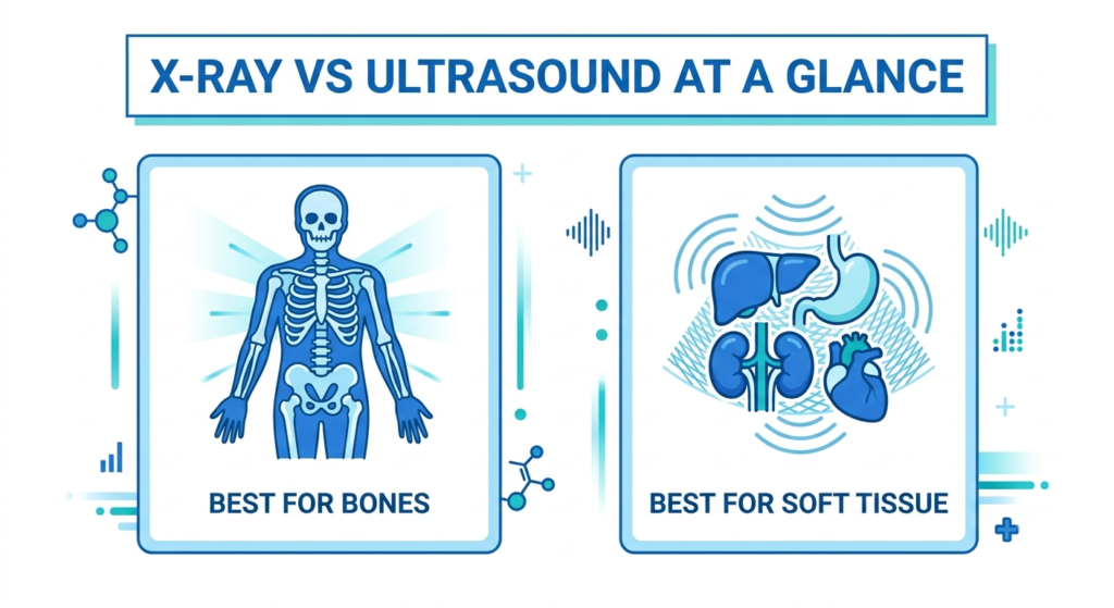
Owners often assume X-rays and ultrasound serve the same purpose, but each modality is suited to different diagnostic questions. Understanding the difference helps explain why your veterinarian might recommend one, the other, or both together.
| Feature | X-Ray (Radiograph) | Ultrasound |
|---|---|---|
| Best for | Bones, teeth, chest cavity, dense structures | Soft tissue, organs, fluid, blood flow |
| Image type | Static, two-dimensional | Real-time, moving image |
| Radiation | Low-dose | None |
| Common uses | Fractures, arthritis, foreign objects, heart size | Organ evaluation, pregnancy, masses, guided sampling |
| Sedation | Rarely needed | Rarely needed |
In many cases, a veterinarian uses both together. A dog with abdominal pain, for example, might receive an X-ray to rule out an obstruction and a follow-up ultrasound to examine organ texture and detect fluid that an X-ray cannot show.
What a Pet Ultrasound Can Reveal
Abdominal Organs and Soft Tissue
Abdominal ultrasound is the most common application, allowing a veterinarian to examine the liver, kidneys, spleen, bladder, and intestines for size, shape, and internal texture. This can reveal masses, cysts, inflammation, thickened intestinal walls, bladder stones, or signs of organ disease that would not be visible through a physical exam.
Pregnancy Confirmation
Ultrasound is the most reliable way to confirm pregnancy in dogs and cats and to check on fetal viability. Because the images update in real time, a veterinarian can observe fetal heartbeats directly, something that cannot be assessed through any other routine diagnostic tool.
Heart Function
An ultrasound of the heart, called an echocardiogram, allows veterinarians to observe the heart’s chambers, valves, and overall function while it is actively beating. This is particularly useful for pets with a heart murmur, an irregular heartbeat, or symptoms like coughing or exercise intolerance that could point to cardiac disease.
Guided Sampling and Biopsies
Because ultrasound provides a live image, it can also guide a veterinarian’s needle precisely to a mass, cyst, or fluid pocket for sampling. This ultrasound-guided approach is far less invasive than exploratory surgery and often provides diagnostic answers with minimal recovery time for your pet.
When Your Vet Might Recommend an Ultrasound for Your Dog or Cat
Veterinarians typically recommend ultrasound imaging in the following situations:
- Unexplained vomiting, diarrhea, or abdominal pain lasting more than a day or two
- Abnormal bloodwork results suggesting organ dysfunction
- A palpable mass or lump detected during a physical exam
- Suspected pregnancy or monitoring of a known pregnancy
- A newly detected heart murmur or irregular heart rhythm
- Straining to urinate or blood in the urine, which may indicate bladder stones
- Trauma from an accident, where internal bleeding or organ injury is a concern
- Monitoring a previously diagnosed condition over time
What Happens During a Pet Ultrasound
A typical ultrasound appointment follows a consistent process from start to finish.
- Preparation. A small area of fur, usually on the abdomen, is gently clipped to allow the probe full contact with the skin. Some pets may be asked to fast beforehand if the scan involves the digestive tract.
- Positioning. Your pet lies on their side or back on a padded table. Most pets tolerate this well with gentle handling and no restraint beyond a technician holding them steady.
- Gel application. A small amount of conductive gel is applied to help the sound waves transmit clearly between the probe and the skin.
- Scanning. The veterinarian moves the probe slowly over the area being examined, watching the real-time image on the monitor and adjusting position to get clear views of each organ.
- Review and discussion. Once the scan is complete, your veterinarian reviews the findings with you, explains what was observed, and discusses next steps if anything abnormal was detected.
The full appointment typically takes between twenty and forty-five minutes, depending on how many organs or areas need to be evaluated.
Is Sedation Needed for a Pet Ultrasound?
Most pets tolerate ultrasound scans well without sedation because the procedure is painless and non-invasive. Gentle restraint is usually sufficient. However, pets who are highly anxious, in pain, or need to remain very still for a detailed cardiac scan may benefit from light sedation. This decision is always made individually, based on your pet’s temperament and the specific area being examined, with comfort as the top priority.
How to Prepare Your Pet for an Ultrasound Appointment
A little preparation goes a long way toward making the visit smooth for both you and your pet. Keep the following in mind before your scheduled ultrasound:
- Follow fasting instructions carefully. If your veterinary team recommends withholding food for a set number of hours before an abdominal scan, stick to that window closely, since food and gas in the digestive tract can obscure the view of nearby organs.
- Bring a calm, familiar item. A favorite blanket or towel from home can help an anxious pet settle more easily on the exam table.
- Avoid a long walk right before the appointment if the bladder needs to be full for the scan. Your veterinary team will let you know if this applies to your pet’s specific procedure.
- Ask questions ahead of time. If you are unsure whether your pet’s current medications should be given as usual, confirm with the clinic before the appointment rather than guessing.
- Plan for a little extra time. While the scan itself is usually quick, allow time for check-in, positioning, and a follow-up conversation with your veterinarian about the findings.
Most pets go home the same day with no restrictions, since ultrasound involves no radiation and rarely requires sedation.
Real Case: How Imaging Guided a Trauma Diagnosis
Ultrasound plays a critical role beyond routine wellness screening, particularly in emergency and trauma situations. When a pet arrives after a fall, a vehicle accident, or another traumatic event, internal injuries are not always visible from the outside. In these cases, ultrasound allows the veterinary team to check for internal bleeding, fluid accumulation, or organ damage within minutes, information that directly shapes whether a pet needs immediate surgical intervention or can be stabilized and monitored.
This kind of rapid, real-time imaging is one of the reasons on-site diagnostic capability matters so much during emergencies. You can read more about how imaging supports surgical decision-making in the hospital’s overview of common veterinary surgeries for cats and dogs, which explains how pre-surgical imaging shapes treatment plans for a range of conditions.
Ultrasound as Part of a Complete Diagnostic Picture
Ultrasound findings are rarely used in isolation. Veterinarians typically combine imaging results with pet lab tests such as bloodwork and urinalysis to build a complete understanding of what is happening inside your pet’s body. If an ultrasound reveals a mass or abnormality that requires surgical treatment, those images become an essential reference for planning veterinary surgical procedures with precision. This combined approach, imaging plus lab work plus physical examination, consistently leads to more accurate diagnoses than any single test on its own.
Having imaging equipment available on-site also matters when timing is critical. In emergency vet near me situations, immediate ultrasound access means a pet can be scanned, diagnosed, and treated in a single visit rather than waiting for a referral appointment elsewhere.
According to the Merck Veterinary Manual, ultrasound has become one of the most widely used diagnostic imaging tools in veterinary medicine because it provides detailed soft-tissue information without the radiation exposure associated with other imaging methods. For more complex or specialized cases, veterinarians may also draw on resources from the American College of Veterinary Radiology, the specialty organization responsible for training and certifying veterinary radiologists, or academic imaging programs such as the one at Cornell University College of Veterinary Medicine, which represent the advanced end of veterinary diagnostic imaging.
Frequently Asked Questions
-
Is a pet ultrasound painful for dogs or cats?
No, pet ultrasound is completely painless and non-invasive. The procedure uses sound waves rather than needles or radiation, and most pets simply feel the gentle pressure of the probe against their skin along with the coolness of the conductive gel. Most animals tolerate the scan calmly, especially with gentle handling and a quiet exam room.
-
How long does a pet ultrasound take?
A standard ultrasound appointment usually takes between twenty and forty-five minutes, depending on how many organs are being examined and whether guided sampling is also performed. Cardiac ultrasounds, which require detailed views of the heart’s chambers and valves, can sometimes take a bit longer than a routine abdominal scan.
-
Does my pet need to fast before an ultrasound?
Fasting is often recommended before an abdominal ultrasound because food and gas in the stomach and intestines can make it harder to see other organs clearly. Your veterinary team will give you specific instructions based on the type of scan being performed, so always confirm before the appointment rather than assuming.
-
What conditions can ultrasound detect that X-rays cannot?
Ultrasound is far better than X-rays at evaluating soft-tissue detail, including organ texture, fluid accumulation, blood flow, and early-stage masses that have not yet changed an organ’s overall shape. It also allows real-time observation of movement, such as a beating heart or fetal activity, which a static X-ray image cannot capture.
-
When should I ask my vet about scheduling an ultrasound?
You should bring up ultrasound imaging if your pet has ongoing vomiting or diarrhea, unexplained weight loss, abnormal bloodwork, a new lump, difficulty urinating, or a suspected pregnancy. Your veterinarian will determine whether ultrasound, X-ray, or a combination of both is the right next step based on your pet’s specific symptoms.
-
Can ultrasound results lead directly to treatment decisions?
Yes. Ultrasound findings often guide immediate next steps, whether that means starting medication, scheduling a biopsy, or moving forward with surgery. Because the images are available right away during the appointment, your veterinarian can often discuss findings and a treatment plan with you the same day rather than waiting on results from an outside facility.
A pet ultrasound offers a clear window into what physical exams and bloodwork alone cannot show, often turning uncertainty into an answer within the same visit.